HKUST Engineering Researchers Develop GenAI Framework for Misalignment-Resistant Virtual Staining to Accelerate Histopathology Workflows
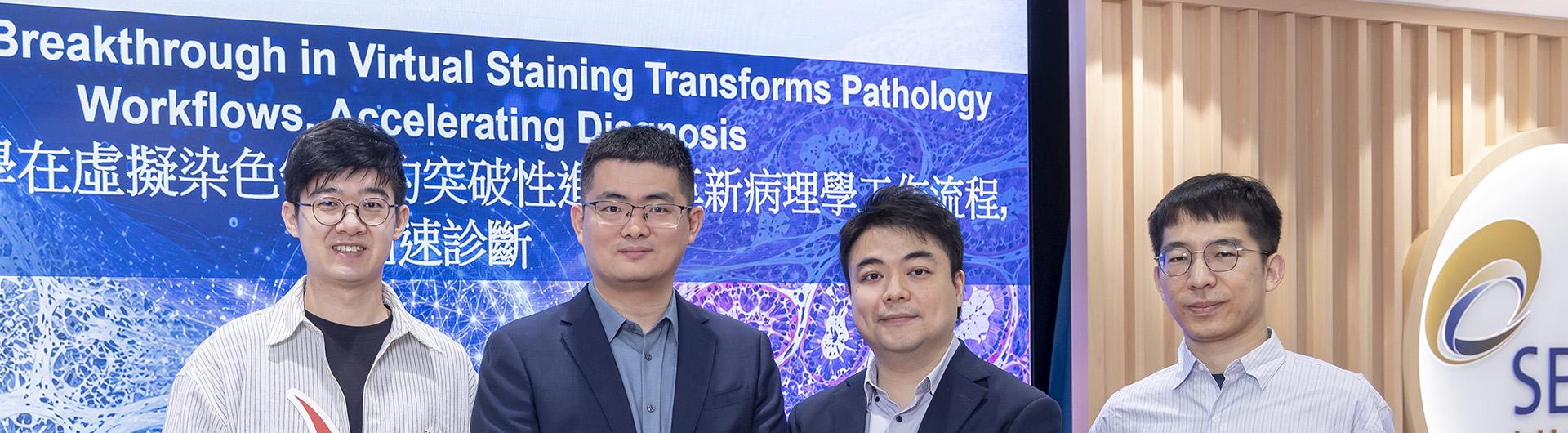
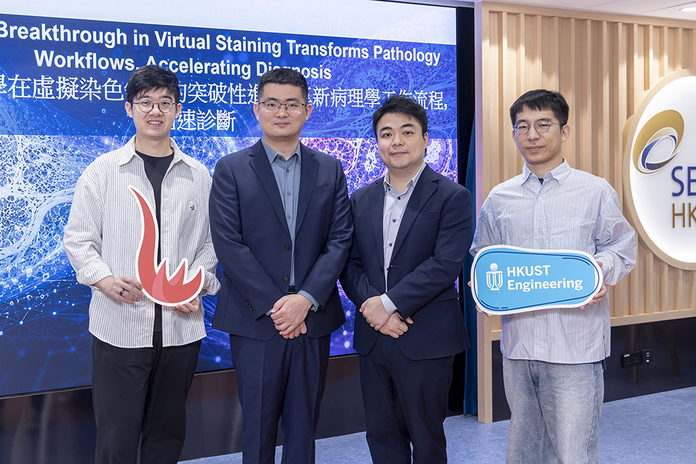
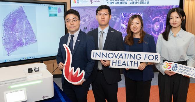
Prof. Hao CHEN, Assistant Professor in the Department of Computer Science and Engineering and the Department of Chemical and Biological Engineering, Director of the Collaborative Center for Medical and Engineering Innovation and SmartX Lab, has developed, in collaboration with Prof. Terence WONG, Associate Head and Associate Professor in the Department of Chemical and Biological Engineering and Associate Director of the Collaborative Center for Medical and Engineering Innovation, along with researchers from the Southern Medical University in Guangzhou, The Chinese University of Hong Kong, and other collaborative partners, a GenAI framework for misalignment resistant virtual staining.
This successful project was also made possible by the dedicated work of co-first authors Jiabo MA and Wenqiang LI, who are both PhD students in the Department of Computer Science and Engineering at HKUST and members of Prof. Chen’s research team.
The Problem with Traditional Testing
When doctors suspect a patient has a disease like cancer, they historically have had to take a small tissue sample (a biopsy) and use chemicals to stain it so they can see the cells clearly under a microscope. This traditional method is slow, requires a lot of manual labor, and can quickly use up tiny, precious tissue samples.
While scientists have previously tried using AI to "virtually" stain these tissue samples digitally, they struggled because the training images were often slightly stretched, bent, or misaligned due to how delicate tissue is handled.
How the New AI Works
To solve this, the research team created a smart AI framework called Decoupled Generation and Registration (DGR). This system is unique because it separates the process of coloring the digital image from the process of fixing any physical stretching or shifting in the tissue.
In testing, this new AI performed incredibly well:
- True-to-life quality: When professional pathologists were asked to tell the difference between real chemically stained tissues and the AI's virtual stains, they guessed incorrectly about half the time—meaning the AI images looked almost identical to real ones.
- Saving tissue and time: This technology allows doctors to get the highly detailed colored images they need much faster, without destroying or running out of the patient's original tissue sample.
- Better cancer detection: When combined with existing medical tools, these high-quality virtual stains helped AI systems do a better job at classifying colon polyps and stomach cancer.
This breakthrough represents a major step forward in making medical diagnoses faster, cheaper, and much easier on patients by preserving their tissue samples.
(This news was originally published by the HKUST School of Engineering here.)